What Are The Advantages Of Ultrasound?

Many people often think about expectant women whenever they hear the expression ‘ultrasound’. Though, even though one of the most predominant applications of ultrasound scanning is fetal imaging, this investigative tool built by Ultrasound Machine Manufacturers has been applied in a wide diversity of use cases in recent times. Among investigative testing techniques, ultrasound imaging is the favored choice for most doctors and physical therapists.
A problem-solving ultrasound unit can offer valued insight into the best treatment or medical procedures to efficiently challenge various health settings, making it an indispensable tool.
Summary of the technology
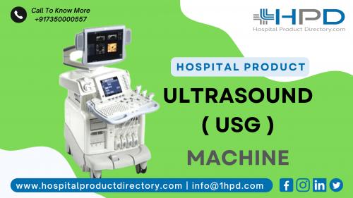
An ultrasound machine made by Ultrasound Machine Manufacturers in India uses sound waves to generate an image of the activities that occur in the human body. A transducer is used to produce high-frequency sound, which is frequently not within human hearing range. The sound reverberations are then logged as the ultrasonic waves bounce back. With this real-time data shown on a computer screen, the size, steadiness, and shape of soft tissues and organs within the body can be regulated. Typically, technicians (sonographers) undergo special drills to conduct imaging procedures. A doctor or radiologist is accountable for construing the resulting ultrasound imaging results. Numerous health circumstances can be identified and treated with this technology.
How is Ultrasound Technology Useful?
Ultrasonography offers many advantages, which is why it has emerged as one of the most effective apparatuses for the analysis of various health conditions and succeeding treatment of them.
Below are some of the main advantages and details of its prevalence.
Safety
No Ionizing Radioactivity: The main advantage of ultrasound imaging is that it uses ultrasonic sound waves to create pictures.
Ultrasound methods differ from other imaging procedures, as no radioactivity is used. As a consequence, any hostile patient response usually produced by radioactivity exposure is avoided.
Other imaging examinations often require substances known as contrast agents. These contrast agents support emphasizing precise parts of the body with issues during diagnostic imaging. Patients are typically administered the agents by oral medicines or injections in blood circulation pathways.
Many people suffer sensitive responses to these substances. Similar contrast agents for ultrasound imaging are not obligatory in most cases, thus safeguarding patient security.
Non-invasive Approach
Ultrasound examinations do not require aggressive procedures.
Technicians only require to place the fitting acoustic transducers in direct contact with the skin over the precise areas that require visualization.
For instance, to check a patient’s thyroid gland, the probe is positioned on the patient’s neck. For expectant women, it is positioned on the stomach.
Trouble-free
Investigative ultrasound approaches are generally painless. After all, they do not require inoculations, incisions, or needles. As a consequence, patients dodge postoperative chronic pain or operative complications.
For instance, simply placing a probe on an expectant woman’s stomach produces a clear picture of her unborn baby. Very informal and effortless. This makes an ultrasonography investigation suitable for various applications.
No retrieval time
Typically, non-invasive systems require no recovery period.
Though, invasive approaches need incisions, which require proper management of the cuts post-procedure. Patients have no necessity to worry about this when it comes to an ultrasonography investigation.
Easy to function
Most mechanics find the ultrasound machine built by Ultrasound Machine Manufacturers informal to operate. The entire process is quite humble.
An ultrasound cream is applied to the patient’s skin to stop air pockets from blocking the ultrasonic waves. Then a sonographer presses the handheld probe against the precise area and moves it around to get a clear picture.
Even when clearer pictures are required, a piezoelectric transducer is simply committed to the probe and implanted into a natural cavity in the body. This comfort of use has also contributed to the admiration of ultrasonography.
Convenience and Speed
Ultrasonography sittings are often rapid; most last for just a few minutes.
The most concentrated ultrasound examinations only take up to an hour. This makes it suitable for people with busy calendars.
Clear Pictures
The clear pictures produced using ultrasonography are a huge advantage. They can support doctors to choose the best approach for treatment.
Most ultrasound machines often come with choices that support the production of clear pictures.
Dynamic real-time pictures
Dynamic real-time 3D imaging safeguards technicians can trust high-quality pictures and timely spatial information of the precise area being scanned.
Pictures are shaped as the probe is moved around the perused area.
Displays soft tissues in great detail
Unlike other imaging devices like X-rays used for probing hard tissue, such as bones, ultrasonography is perfect for visualizing soft tissues.
When the ultrasound waves meet tissues with varying thicknesses (e.g., healthy tissue, non-healthy tissue), these reproduce different echo designs, which can then be computed.
What are the dangers of consuming ultrasound?
There are no recognized damaging effects. The technique accepts low-power sound waves and is a respected tool that can be used for many applications. Though, certain restrictions cannot be ignored.
Conclusion:
Ultrasound examinations use high-frequency sound waves to produce pictures of the body. Thanks to their many benefits, ultrasound pictures are used in a wide array of applications. It can also notice injury to veins and display blood flow in blood vessels. With no recognized dangers, the advantages of ultrasonography have been safeguarded that medical professionals can easily endorse it if available. Ultrasonography is also suitable, informal to do, and can be fitted easily into a busy calendar.

Comments