How does a Neuro Microscope help in Image Guided brain Tumor Surgery?

The numerous applications of navigation for neurosurgery have been extensively used and conveyed for almost two decades. Today, doctors are using high-tech technologies that are acquainted with us in the consumer world to help them fight cancer in the operating room. An imperative example of that application is image-guided surgery (IGS) and it aids surgeons to complete harmless and less aggressive measures and eliminate brain tumors that were once measured inoperable due to their scope and/or site.
Similar to a car or mobile Global Positioning System (GPS), image-guided surgery schemes use cameras or electromagnetic fields to seize and communicate the patient’s structure and the surgeon’s exact movements about the patient, to computer screens in the operating room. These sophisticated high-tech systems are used before and during surgery to help position the surgeon with three-dimensional pictures of the patient’s structure including the tumor.
Surgeons and surgical staff can:
Gage the location, scope, and site of a patient’s brain tumor in association with the edifices in the brain and plan the anticipated admission
Plot the site of the craniotomy in association with the brain tumor
The path of the surgical tools in association with the patient’s brain and the tumor itself
Recognize and reason for vagaries in the brain during the process
What is the role of a Neuro Microscope?
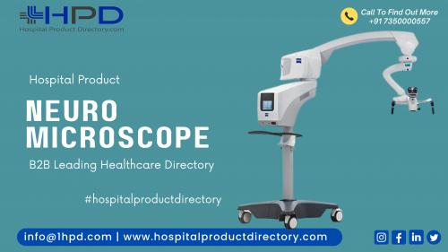
Image-guided surgery offers a road map for surgeons to help plot out the straight and harmless way to access a brain tumor. Since neurosurgeons complete most of their actions while looking through a Neuro microscope made by the Neuro Microscope Manufacturers, microscope addition with image-guided surgery is a very valuable tool used during any brain process. It permits the neurosurgeon to switch between live pictures of the patient as well as pictures taken of the patient pre-surgery. Surgeons continually need to distinguish where they are during surgery. Before the Neuro microscope addition, surgeons had to halt what they were doing and look at another steering screen to find their site. Being able to view steering information through the Neuro Microscope while looking at the patient in real-time saves the surgeon time during surgery, making it a valued tool during surgical treatment.
Characteristically, a surgery that is steered follows several steps:
The patient’s diagnostic pictures like CT or MR are uploaded into the IGS system where the doctor can then generate a strategy for the surgery. This strategy shows a full color, patient-specific 3D model of the brain tumor, the fit tissue, and serious brain constructions near and around the brain tumor like the optic and cranial nerves and useful areas, accountable for hand and leg movement and the brain stem, for instance.
This 3D model, which looks like animatronics of the patient’s brain, is listed to the actual location of the patient on the operating table so that the surgeon can realize or ‘track’ his tools about the patient’s real composition and position themselves on the 3D animatronics shown on the computer screen.
The patient can be followed with different chasing technologies, which may comprise optical or electromagnetic. With the optical technology, the structure needs special reflective indicators on a reference tool, which is positioned close to or onto the patient’s head. These reflective indicators are also situated on the operating instruments and are pursued by an infrared camera, which is linked to the system’s computer.
With all this evidence counting the scope, size, and location of the brain tumor, the surgeon can achieve brain surgery while lessening the risk to critical edifices and fit tissue.
Stages For Using Image-Guided Brain Tumor Surgery
Treatment Arrangement and Patient Groundwork
Your surgeon has uploaded your pre-operative analytical data to the image guidance structure to get a modified 3D model of your skull, brain, and critical structures. With this evidence, the surgeon strategies the surgery by delineating the outlines of your brain tumor, making sure to avoid the fit tissue and any significant structures near the tumor. Once the healing plan is equipped, it is saved on the steering system until being recovered from inside the operating room.
Once you arrive at the hospital, you are given anesthesia as groundwork for surgery. When you are lifeless and before your doctor makes any cuts, the part where the craniotomy will be done will be hairless and will be disemboweled. In most cases, your head will be restrained so that you remain motionless throughout the procedure.
Patient Registering
A special laser indicator permits surgeons to list you to the image-guided surgery system by skimming the exterior of your face without touching it. This ‘surface matching’ procedure is a precise and easy-to-use patient registering method that mechanically computes your cranial construction and brings into line it with your 3D model on the computer screen. After your operating room location has been affiliated with the virtual map shaped by the surgeon before surgery, the IGS system can track your brain and the doctor’s operating instruments throughout the process and virtually in real time.
Craniotomy and Image Guided Brain Tumor Elimination
Founded on the pre-planned steering route, and once you are surgically swathed, your doctor will perform the craniotomy as recognized in the preparation stage and directed by the neuronavigation system. The surgeon can check the pictures on the computer monitor to make sure that the actual cut is in the right place (and at the right approach) as prearranged on the 3D brain typical before surgical treatment.
Once the craniotomy is finished, your surgeon will access the brain tumor according to the pre-planned course, or way, while eventually evading critical areas of the brain. At any time, the neurosurgeon can shadow each of his or her phases on the IGS system monitor. Incorporation and tracking with a neuro microscope made by the Neuro Microscope Manufacturers in India or an ultrasound machine may give your surgeon added visual direction throughout your tumor elimination.
Conclusion
Once the anticipated goal of your surgery is attained—fractional or full brain tumor elimination—the craniotomy is fastened and you are brought to the recovery room. Since image-guided surgery with neuronavigation may decrease the intrusiveness of the surgery, you may likely return home only a few days after hospitalization, and revert to everyday life. Though, this will be contingent upon your surgery, your retrieval, and your physician’s endorsements. Contingent on your tumor, your surgeon may endorse chemotherapy or radiotherapy as a complement to your surgery.
Post Your Ad Here
Comments